
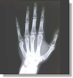
Dayton C. Miller, a physicist at the Case School of Applied Science in Cleveland, did the first experiments using X-rays to observe fractured bones. X-rays had been discovered by Wilhelm Roentgen in 1895. But soon after, Miller perfected the techniques for exposing skeletal images on photographic plates. He decided to use cathode ray tubes built by William Crookes to make some of the first photographic images of concealed objects, including a bullet within a man's limb.
Miller earned his doctorate in astronomy at Princeton University, and then in 1890, he began his work at the Case School of Applied Science as an astronomy teacher. Later, he focused on physics and became the head of the physics department in 1893.
What is an X-Ray?
Light has many different forms, including visible light, and others such as radio waves, microwaves, infrared, ultraviolet, gamma radiation, and X-rays. The energy of the photon indicates its form of light. For example, radio waves have low energy photons. Optical photons, which are the ones we can see, are a million times more energetic than those in a radio wave. And, the energies of X-ray photons can be hundreds to thousands of times greater than those of optical photons.
Did You Know?
- Miller worked with George Crile, leading to use of X-rays in cancer treatment.
- Other Cleveland Clinic scientists made key advances in determining proper doses of x-rays.
- Dayton Miller collected flutes. He is said to have had flutes made by 460 European and American instrument makers, over 130 Native North and South American flutes, and a 17th/18th century jade flute example from China. Miller died in Cleveland in 1941 as he was preparing to move his entire collection of flutes, books, and related materials to the Library of Congress.
Find out more...